
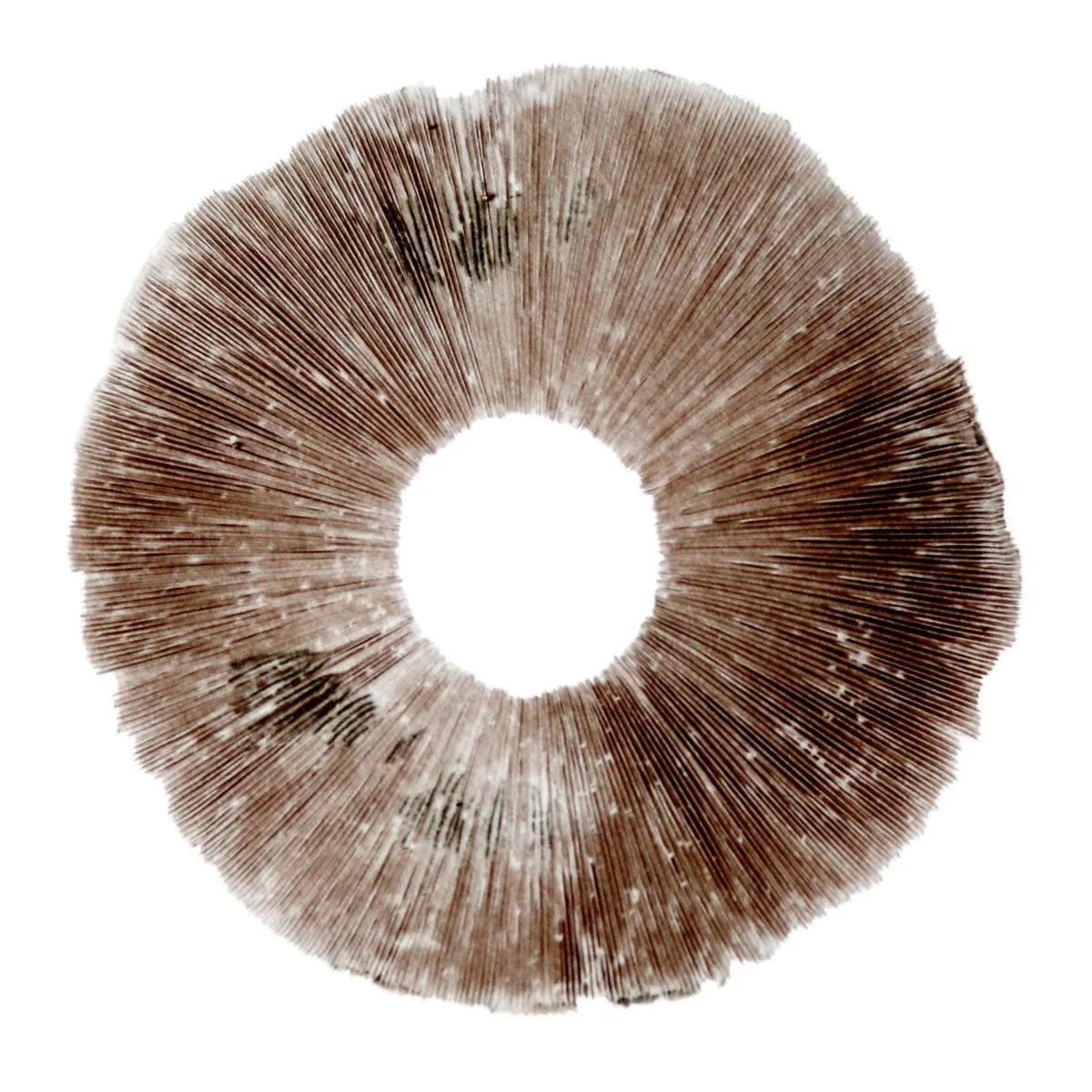
Spore Print Psilocybe cubensis Hawaii (PES)
Buy more, pay less
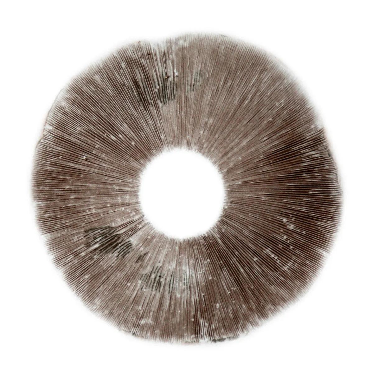
Psilocybe Cubensis Hawaii (PES) Spore Print
The Psilocybe cubensis Hawaii spore print is a single-strain specimen on foil, ready for microscopy study of one of the more enigmatic cubensis varieties in circulation. Despite carrying the name "Hawaii," nobody has definitively traced this strain to the islands — the link comes from Pacific Exotica Spora (PES), a Honolulu-based company that first brought it to market. What we do know: the spore morphology bears a striking resemblance to cubensis Cambodia, which has led plenty of cultivators to speculate they share common genetics. Whether that's true or just convergent evolution in Petri dishes, the Hawaii PES remains a popular and widely studied strain.
What Makes the Hawaii PES Cubensis Spore Print Worth Studying
The Hawaii PES spore print sits in an interesting spot among cubensis varieties — it's a strain with a murky origin story and a dedicated following. Experienced microscopists often note that the spore structure closely mirrors cubensis Cambodia, with dark purple-brown deposits that are dense and well-defined on the foil. That density matters: a good spore print should give you enough material for multiple slides without running thin halfway through your session.
Under the microscope at 400-1000x magnification, cubensis Hawaii spores typically present as subellipsoid, measuring roughly 11-17 x 8-11 micrometres. The cell walls are relatively thick, which makes them forgiving for beginners still getting the hang of staining techniques. If you've worked with Golden Teacher prints before, you'll find the Hawaii PES comparable in spore density but often slightly more uniform in size distribution — fewer outliers, cleaner fields of view.
One honest note: because the Hawaii PES origin is genuinely uncertain, you won't find the same depth of published genetic data as you would for, say, a well-documented B+ or Amazonian isolate. According to research published in Fungal Biology, DNA authentication of Psilocybe species and strains remains an evolving field, with increasing evidence that many commercially traded cubensis varieties share closer genetic relationships than their names suggest (PMC9764976). That's part of what makes comparative microscopy between strains like Hawaii PES and Cambodia genuinely interesting work.
Cubensis Hawaii PES Spore Print Specifications
Every spore print ships on aluminium foil in a sealed, sterile packet. Here's what you're getting:
| Specification | Detail |
|---|---|
| Species | Psilocybe cubensis |
| Strain | Hawaii (PES — Pacific Exotica Spora) |
| Format | Spore print on aluminium foil |
| Spore colour | Dark purple-brown |
| Typical spore dimensions | 11-17 x 8-11 micrometres (subellipsoid) |
| SKU | SH0119 |
| Storage | Cool, dark, dry — sealed until use |
| Shelf life (sealed) | 12-24 months at room temperature |
| Intended use | Microscopy and taxonomy research |
How to Use a Cubensis Spore Print for Microscopy
Working with a spore print is straightforward, but a few details make the difference between a clean slide and a frustrating afternoon. Here's the process we'd follow:
- Prepare your workspace. Wipe down your surface with isopropyl alcohol (70% works well). Have your microscope slides, cover slips, sterile water or staining solution, and a scalpel or inoculation loop ready before you open the print.
- Open the sealed packet carefully. The foil will have a visible deposit of dark purple-brown spores. Avoid breathing directly over the print — not because it's dangerous, but because you'll scatter spores everywhere and thin out your sample.
- Scrape a small quantity of spores onto a clean glass slide using the edge of a sterile scalpel or loop. You need far less than you think — a barely visible amount will give you thousands of individual spores under magnification.
- Add a single drop of sterile water or your preferred mounting medium. For basic observation, water works fine. For detailed morphological study, a drop of Melzer's reagent or cotton blue in lactic acid will highlight cell wall structures.
- Place a cover slip at a 45-degree angle and lower it gently to avoid air bubbles. Trapped air creates dark circles that obscure your field of view.
- Start at 100x magnification to locate spore clusters, then move to 400x for individual spore morphology. At 1000x with oil immersion, you can observe fine details like the germ pore and ornamentation patterns.
- Reseal the remaining print in its foil packet and store in a cool, dark place. A sealed print keeps for 12-24 months at room temperature without significant viability loss.
Storing Your Cubensis Hawaii Spore Print
Spore prints are remarkably shelf-stable compared to syringes or liquid cultures, but they're not indestructible. The enemies are moisture, heat, and UV light — in that order. A sealed print left in a desk drawer at 18-22 degrees Celsius will last 12-24 months without noticeable degradation. We've seen prints stored in fridges (4-8 degrees Celsius) that were still producing clean slides after 3 years, though we wouldn't bank on that as a guarantee.
The one thing that kills prints fast: opening and resealing repeatedly in a humid room. Every time you expose the foil, ambient moisture can settle on the spore deposit and encourage bacterial contamination. If you plan to use the print across multiple sessions, consider cutting the foil into sections on your first opening, resealing each piece individually. A small ziplock bag with a silica gel packet does the job.
Building a strain library? Pair this Hawaii PES print with a cubensis Cambodia spore print for side-by-side comparative microscopy — the morphological similarities make them a natural study pair. If you're new to working with prints, a spore syringe gives you a ready-to-use liquid suspension that's easier to handle on your first few slides.
The Science Behind Psilocybe Cubensis Spores
Psilocybe cubensis is the most widely distributed and studied species in the Psilocybe genus. According to a global species survey published in 2023, psychedelic fungi "have received considerable attention recently due to their promising treatment potential of several psychiatric disorders" (PMC10238702). The psychotropic compounds — psilocybin and psilocin — were first isolated and characterised in the late 1950s by Albert Hofmann, and research into their pharmacology has accelerated dramatically since 2016 (PMC5220184).
From a microscopy perspective, cubensis spores are among the easiest to work with in the Psilocybe genus. The dark purple-brown spore deposit is dense, the individual spores are large enough to study without oil immersion (though it helps), and the subellipsoid shape with a distinct germ pore makes identification straightforward. Research published in 2020 noted that studies "support safe use of mushrooms under controlled conditions" while emphasising the importance of proper identification and characterisation (PMC7749179). Accurate spore identification through microscopy is a foundational skill in that process.
What to Expect from This Spore Print
Let's be straight about what you're getting: a single spore print on foil, sealed in a sterile packet. It's not a grow kit, it's not a syringe, and it doesn't come with slides or mounting medium. What it does give you is a generous deposit of Psilocybe cubensis Hawaii (PES) spores — enough material for dozens of microscopy slides if you're conservative with your scraping.
The print arrives dry and sealed. You should see a clear, dark deposit on the foil — if the deposit looks patchy or thin, that's unusual and worth flagging with our customer service team. A good cubensis print looks like a solid stamp of dark purple-brown, sometimes with visible gill-line patterns from the cap that produced it. Those gill lines are actually useful landmarks when you're trying to scrape from a consistent area of the deposit.
Frequently Asked Questions
What magnification do I need to study cubensis Hawaii PES spores?
Start at 100x to find spore clusters, then move to 400x for individual spore morphology. Oil immersion at 1000x reveals fine details like germ pore structure and wall ornamentation. A basic compound microscope with 400x capability covers most identification work.
How long does a Psilocybe cubensis spore print last in storage?
Sealed and stored in a cool, dark place at 18-22 degrees Celsius, a cubensis spore print remains viable for 12-24 months. Refrigerated prints (4-8 degrees Celsius) can last even longer — we've seen usable prints after 3 years in the fridge, though results vary.
Is cubensis Hawaii PES the same as cubensis Cambodia?
Nobody knows for certain. The spore morphology is strikingly similar, and many cultivators suspect they share common genetics. Without published molecular data comparing the two directly, the question remains open — which is exactly why having both prints for comparative microscopy is worthwhile.
What does PES stand for in Hawaii PES?
PES stands for Pacific Exotica Spora, the Honolulu-based company that first commercialised this strain. Despite the Hawaiian branding, the actual geographic origin of the strain is unknown. The PES designation simply identifies the original vendor.
How many microscopy slides can I make from one spore print?
A standard cubensis spore print contains millions of individual spores. With conservative scraping — using just the tip of a scalpel or inoculation loop — you can prepare 30-50 slides from a single print before the deposit starts thinning noticeably.
Do I need staining solution to view cubensis spores?
No. Cubensis spores are naturally pigmented (dark purple-brown) and visible in a simple water mount at 400x. Staining with Melzer's reagent or cotton blue in lactic acid enhances cell wall detail but isn't required for basic identification.
What's the difference between a spore print and a spore syringe?
A spore print is a dry deposit on foil — shelf-stable, compact, and gives you control over how much material you use per slide. A spore syringe is a liquid suspension in sterile water, ready to drop directly onto a slide. Prints last longer in storage; syringes are more convenient for quick prep.
Last updated: April 2026
Related products
You might also like
Medical disclaimer. This content is for informational purposes only and does not constitute medical advice. Consult a qualified healthcare provider before use of any substance.